Loading...

Table of Content

    31 August 2024, Volume 5 Issue 4
    Invited Review
    Early human embryo development and stem cells
    Zongyong AI, Chengting ZHANG, Baohua NIU, Yu YIN, Jie YANG, Tianqing LI
    2024, 5(4):  700-718.  doi:10.12211/2096-8280.2023-094
    Asbtract ( 360 )   HTML ( 27)   PDF (1982KB) ( 404 )  
    Figures and Tables | References | Related Articles | Metrics

    The early development of the human embryo includes three important stages: ①the pre-implantation stage from the zygote to the late blastocyst; ②the peri-implantation stage from late blastocyst to pre-gastrulation embryo; and ③the most mysterious post-gastrulation stage from gastrulation to early organogenesis. The latter two stages are collectively referred to as the early post-implantation developmental stage. During pregnancy, infertility (implantation failure or miscarriage) and birth defects of the fetus are largely due to abnormalities in human early postimplantation development. Human early postimplantation embryo, due to its small size and location in the mother’s uterus, is difficult to observe and study. Therefore, the embryonic development process at this stage has been in a black box state for a long time. In recent years, with the emergence of single-cell omics technology and extended in vitro culture system of human blastocysts, as well as the rapid development in the fields of embryonic and extraembryonic stem cells, organoids and embryoids, the mystery of the human early postimplantation development is gradually being lifted. In order to help understand the mysteries of early human embryonic development, this review primarily introduces the lineage diversification, key developmental events and known developmental principles during early human embryogenesis; summarizes recent progress in the research on human embryonic and extraembryonic stem cells (including totipotent stem cells, embryonic stem cells, trophoblast stem cells, primitive endoderm stem cells and extraembryonic mesoderm cells); presents the effects of cell communication, lineage interaction, signal gradient, adhesion molecules, biomechanics, and extracellular matrix on cell sorting, migration rearrangement and self-organization in embryoids and organoids; reviews the current research status of human early post-implantation embryogenesis, stem cell-based embryo models and organoids; and finally proposes the prospects and possible solutions to the problems and challenges existing in the research of human early post-implantation development using stem cell-derived embryo models or organoids.

    Advances in the development of human embryo models
    Bowen HU, Jiaping TAN, Xiaodong LIU
    2024, 5(4):  719-733.  doi:10.12211/2096-8280.2024-010
    Asbtract ( 624 )   HTML ( 20)   PDF (1524KB) ( 302 )  
    Figures and Tables | References | Related Articles | Metrics

    Human early embryonic development is critical for a healthy fetus birth. However, the specific molecular regulatory mechanisms of lineage development, cell fate decisions and embryonic patterning are still shrouded in mystery. Our knowledge about early human embryogenesis has been greatly improved with the recent progress in in vitro culture conditions for human blastocysts and the advancements in omics technology. However, ethical and technological challenges continue to pose obstacles in these studies. With the rapid development of human pluripotent stem cells, they can be coaxed to form embryo-like structures that mimic early human embryonic development in vitro, termed “embryo models”. Interest in human embryonic development has been reinvigorated with the continuing advances in this area. Human embryo models can be divided into two categories "non-integrated embryo models" and “integrated embryo models” according to the different cellular components they possessed. Integrated embryo models represent the embryo-like structures containing both embryonic and extra-embryonic cell types, including blastoids, human extra-embryoids (hEEs), E-assembloids, stem-cell-derived synthetic whole embryo models (SEMs), peri-gastruloids, bilaminiods and heX-embryoids. While non-integrated embryo models sometimes lack the extra-embryonic tissues, including embryoid bodies (EBs), gastruloids, micro-patterned colonies, post-implantation amniotic sac embryoids (PASE). Besides, non-human primate cynomolgus monkey models have greatly expanded our knowledge towards human developmental biology. In this review, we summarized recent human stem cell-based non-integrated and integrated embryo models, and pointed out the technical challenges remained with proposed future directions. These findings lay an important foundation for understanding early human embryonic development, promoting research into human stem cells and their application, as well as preventing and treating early pregnancy loss or congenital diseases. On the other hand, embryo-like structures derived from human stem cell-based integrated embryo models, although do not fully, almost recapitulate the key events and structural organization of the in vivo counterparts, thus eliciting a serious compact on traditional ethical and practical concerns. In the future, non-integrated human embryo models and non-human primate models, which pose fewer ethical challenges, may provide a straightforward way to study the human early embryonic development.

    Stem cell-based synthetic development: cellular components, embryonic models, and engineering approaches
    Yizhao HAN, Jia GUO, Yue SHAO
    2024, 5(4):  734-753.  doi:10.12211/2096-8280.2023-100
    Asbtract ( 188 )   HTML ( 10)   PDF (2285KB) ( 165 )  
    Figures and Tables | References | Related Articles | Metrics

    Over the past century, the scientific foundation of embryonic development has primarily relied upon meticulous examination of developmental processes in model organisms. However, investigating the development of mammals has presented numerous challenges, including interspecies disparities, ethical considerations, and technical constraints. With the rapid advancement of stem cell technology, researchers have endeavored to overcome these obstacles by harnessing the potential of stem cells to generate sophisticated invitro embryo models. The rapid advancement of stem cell technology has revolutionized our approach to study embryonic development. While the ability of current embryo models to fully simulate the authentic developmental process is yet to be verified, they undeniably present new possibilities for developmental biology research. This review primarily focuses on mouse and human, summarizing the types of stem cells used in constructing embryo models and elucidating the roles and importance of different stem cells in simulating developmental processes. This review systematically presents and dissects crucial events and spatiotemporal dynamics in the embryonic development of both mice and humans across various stages. We thoroughly discuss the remarkable milestones achieved by existing embryo models, explore methods for evaluating the biomimicry of these models, and highlight the crucial role of bioengineering methods in embryo model development. The pivotal role of bioengineering in advancing embryonic model development is underscored, emphasizing its indispensable contribution to providing the requisite technical scaffolding for the realization of instruct multicellular induced self-organization with high-level spatiotemporal orders. Additionally, we provide perspectives for the optimization and progressive refinement of embryo models, so as to improve their relevance and applicability. In summary, engineered advances in stem cell-based synthetic development could not only improve our understanding of the inherent complexities of embryos, but also hold the potential for applications in disease research, drug screening, reproductive medicine, toxicological assessments, and other related fields, thereby opening new avenues for both fundamental and translational research.

    Research progress of brain organoids in regenerative medicine
    Yuan HONG, Yan LIU
    2024, 5(4):  754-769.  doi:10.12211/2096-8280.2023-102
    Asbtract ( 219 )   HTML ( 18)   PDF (1257KB) ( 219 )  
    Figures and Tables | References | Related Articles | Metrics

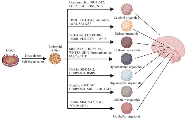
    The brain, as the epicenter of human intelligence, sensation, and motor coordination, represents the pinnacle of biological complexity. Despite its critical role, the availability of live human brain tissue for research is fraught with challenges, impeding advancements in our understanding of the nervous system. Brain organoids are sophisticated three-dimensional cultures derived from human pluripotent stem cells that emulate the diverse cellular composition, structural intricacies, and functional attributes of the human brain. These organoids eclipse traditional two-dimensional cultures and animal models in mirroring the brain’s spatial organization and cellular interplay, bolstered by a genetic congruence with their human counterparts. This congruence renders them particularly adept at modeling neuropsychiatric conditions and pioneering cell-based therapeutic interventions. Regenerative medicine, a confluence of engineering and biological sciences, endeavors to restore tissues and organs compromised by aging, disease, or trauma. However, the field grapples with limitations stemming from the scarcity of samples and ethical quandaries. Brain organoid technology emerges as a formidable asset in this domain, offering expansive potential and profound implications for scientific inquiry. Recent strides have seen the successful assembly of organoid models representing various brain regions through the application of tissue engineering and directed differentiation. These models hold promise for simulating neuropathological states and facilitating tissue repair. This article meticulously surveys the cutting-edge methodologies for constructing organoids specific to brain regions such as the cerebral cortex, hippocampus, striatum, midbrain, thalamus, hypothalamus, cerebellum, and retina. It delineates the principal applications of brain organoids in regenerative medicine, encompassing injury simulation, exploration of inter-regional and multi-lineage cellular dynamics, drug efficacy and toxicity assessments, and the potential for organoid transplantation. Furthermore, the review addresses the prevailing obstacles in the application of brain organoids, notably their pronounced variability, absence of vascularization, and developmental immaturity. In essence, this review seeks to illuminate the organoid generation techniques tailored to discrete brain territories and their significance in regenerative medicine’s landscape. By probing into research poised to surmount the limitations of current models, it aspires to broaden the horizons for brain organoids in both foundational research and clinical applications.
    Advancements in testicular organoids for in vitro spermatogenesis
    Bohang ZHANG, Xiaoxuan QI, Yan YUAN
    2024, 5(4):  770-781.  doi:10.12211/2096-8280.2023-095
    Asbtract ( 349 )   HTML ( 17)   PDF (1768KB) ( 618 )  
    Figures and Tables | References | Related Articles | Metrics

    As the global issue of infertility continues to escalate, particularly with the increasing incidence of male infertility, research in testicular organoids offers new hope and strategies in this field. This review comprehensively discusses the application of testicular organoids in simulating the natural sperm-producing environment, delving into the mechanisms of spermatogenesis, and addressing challenges in male reproductive health. Firstly, we introduce the cellular composition, physiological functions, and the complete process of spermatogenesis within the testicular organ, emphasizing the crucial role of the testicular somatic cell microenvironment in normal testicular development and sperm production. Subsequently, we provide a comprehensive review of the construction of in vitro spermatogenesis systems and the associated research progress through techniques such as testicular tissue culture and reconstruction of testicular organoids in vivo. Moreover, testicular organoids, as a system mimicking spermatogenesis environments in vitro, exhibit significant potential in exploring molecular mechanisms, drug screening and toxicity assessment, as well as preserving and restoring male fertility. Finally, we discuss the limitations of current research in the field of testicular organoids and future research directions. Challenges include accurately simulating the physiological processes of the testis in vitro and improving the quality of sperm obtained in vitro for clinical applications. Future research directions involve delving into the complex interactions between germ cells and somatic cells, aiming to better simulate the testicular microenvironment in vitro, and striving towards safe and effective translation of these research findings into clinical applications for treating male infertility. Additionally, we should ensure that the genetic stability and functionality of germ cells cultured in vitro meet the requirements for clinical applications, and pay attention to the relevant ethical issues. Despite the complexity of the testicular microenvironment and the challenges in fully replicating human spermatogenesis in vitro, the ongoing development in the field of testicular organoids holds promise for providing novel solutions in clinical reproductive medicine and male health research.

    Advances in synthetic biology for engineering stem cell
    Bingyu CAI, Xiangtian TAN, Wei LI
    2024, 5(4):  782-794.  doi:10.12211/2096-8280.2023-101
    Asbtract ( 368 )   HTML ( 44)   PDF (1428KB) ( 257 )  
    Figures and Tables | References | Related Articles | Metrics

    Pluripotent stem cells are characterized by self-renewal and multi-differentiation potential, which can be used to reverse structurally dysfunctional tissues and organs back to a structurally and functionally intact state of health through repair, replacement, or in-situ regeneration of new cells, tissues, and even organs. Cells or multicellular systems derived from pluripotent stem cell differentiation, especially organoids, have great potential for application in regenerative medicine. However, the clinical application of stem cell-related therapies is still in its infancy, and the current challenges to the clinical translation of stem cells include the tumorigenicity, heterogeneity, and immunogenicity of stem cell derivatives. Synthetic biology, with its “top-down” design concept and powerful toolkit including synthetic receptors and gene circuits, allows for the rational assembly of standardized modules. With the rapid development of gene editing technology and the deepening of cell biology research, the engineering object of synthetic biology has shifted from lower model organisms such as Escherichia coli or Saccharomyces cerevisiae to mammalian cells. On the one hand, “top-down” design strategies can engineer stem cells by giving them new functions, and on the other hand, the acquisition of new phenotypes by stem cells can test known gene functions and improve understanding of cell biology. Therefore, the application of these synthetic biology tools to stem cell engineering provides new strategies and platforms for relevant cell therapies or organ transplantation. It offers potential advantages in precise control of cell fate, regulation of cell communication, optimization of organoid structure and function, and monitoring and elimination of tumorigenic cells. These synthetic biology tools have provided new strategies and platforms for the engineering and reprogramming of stem cells, offering the potential to address current challenges in the clinical application of stem cells. They are expected to drive further advancements in regenerative medicine and ultimately achieve the core goal of regenerative medicine, which is organ regeneration.

    Integrated development of organoid technology and synthetic biology
    Ziling CHEN, Yangfei XIANG
    2024, 5(4):  795-812.  doi:10.12211/2096-8280.2023-106
    Asbtract ( 395 )   HTML ( 17)   PDF (1562KB) ( 369 )  
    Figures and Tables | References | Related Articles | Metrics

    Organoids, derived from adult or pluripotent stem cells through invitro differentiation, can recapitulate the cellular diversity, spatial organization, and physiological functions of invivo organs or tissues. The development of organoids has facilitated progress in developmental biology, genetics, pathology, and others. As an emerging interdisciplinary field guided by engineering principles, synthetic biology aims to design, modify, and construct biological components and systems with certain specifically designed functions through engineering and modular approaches. The invitro construction of organoids currently faces several challenges, including high cost, significant heterogeneity, and low throughput, which become more prominent when building complex organoid models. As a burgeoning field in recent years, synthetic biology has excellent potential to expand its applications and research directions. The optimization strategy of organoid construction has become intricately intertwined with the principles of synthetic biology in recent years. Simultaneously, the advancement of synthetic biology and its associated methodologies has propelled the progression of organoid technology. This review provides an overview of the historical developments and current challenges of organoids and synthetic biology while exploring the disparities and interconnections among these fields regarding research concepts and methods. Particularly, we will provide an overview of current design strategies for optimizing organoids and explore the fundamental applications of synthetic biology strategies in this context. Furthermore, we will examine the emerging role of synthetic biology tools in enhancing spatiotemporal fate regulation, structural self-organization, and functional capabilities of organoids. Lastly, we will discuss how derivative research based on organoid platforms contributes to advancing synthetic biology investigations. Overall, this review aims to elucidate the profoundly synergistic and mutually beneficial relationship between the rapidly evolving field of synthetic biology and organoid technology. By delving deep into the interconnectedness of these two disciplines, our objective is to facilitate further exploration of their potential integration in future research endeavors. Additionally, we seek to unravel feasible application scenarios that can harness the combined power of these two fields to bring about potential advancements in biomedical and life science.

    Advances in the application of liver on a chip in biomedical research
    Xiyue CHEN, Yaqing WANG, Fang BAO, Jianhua QIN
    2024, 5(4):  813-830.  doi:10.12211/2096-8280.2024-064
    Asbtract ( 84 )   HTML ( 2)   PDF (2016KB) ( 93 )  
    Figures and Tables | References | Related Articles | Metrics

    The liver plays an important role in maintaining normal physiological activities of the human body. It has a complex structure and multiple functions, including blood glucose regulation, protein synthesis, detoxification and drug metabolism. Although traditional two-dimensional cell culture and animal models have been used to study liver physiology or pathology, there are still some limitations in truly reflecting the microenvironment of human liver and its response to drugs. The development of liver models in vitro is essential for disease research and effective drug testing. Organ-on-a-chip is a groundbreaking technology that has emerged in recent years by merging engineering and biological approaches. It can replicate the essential structural and functional features of human tissues and organs in vitro. Recently, in vitro liver tissue models created with organ chips have shown an impressive ability to closely mimic the liver tissue microenvironment and offer high-throughput capabilities. These models have been extensively applied in liver regenerative medicine, disease research, and drug testing, highlighting their significant potential in the biomedical field. Therefore, in this paper, we provide a comprehensive overview of the limitations inherent in traditional liver in vitro models, particularly in their ability to replicate complex physiological microenvironments and accurately reproduce liver-specific functions. It delineates the design strategies, technical characteristics, and research advancements associated with novel liver in vitro models, with a particular emphasis on organ-on-a-chip technologies. The discussion focuses on the key elements crucial for the biomimetic construction of liver organ-on-a-chip systems and the simulation of liver tissue microenvironments. These elements include the integration of multicellular components, the replication of liver sinusoid and lobule structures, the establishment of biochemical factor gradients, and the incorporation of fluid dynamics. Moreover, it provides an outlook on the future development of highly physiologically relevant liver organ-on-a-chip and microphysiological systems, considering the integration of advanced techniques such as organoids, biomaterials, and gene editing.

    Advances in placenta-on-a-chip for reproductive medicine research
    Rongkai CAO, Jianhua QIN, Yaqing WANG
    2024, 5(4):  831-850.  doi:10.12211/2096-8280.2024-044
    Asbtract ( 137 )   HTML ( 15)   PDF (2513KB) ( 258 )  
    Figures and Tables | References | Related Articles | Metrics

    The placenta is an indispensable organ that connects the mother and fetus, playing various roles during pregnancy such as material exchange, hormone secretion, immune regulation, and barrier defense, which are crucial for maintaining normal fetal development. The placental barrier, composed of multiply layers including trophoblasts, basal lamina and fetal capillaries, plays a crucial role in protecting fetus from direct exposure to xenobiotics. Dysfunction of the placenta can lead to various pregnancy complications, such as preeclampsia, fetal growth restriction, and preterm birth, increasing both maternal and fetal morbidity and mortality rates. Although conventional two-dimensional (2D) cell cultures and animal models have been utilized to study placental physiology and pathology, they still have limitations, such as aberrant cell phenotypes and immature functions in 2D cultures as well as inter-species disparities in animal models. Organ-on-a-chip is a microfluidic cell culture device that allows to mimic the tissue microenvironment by control of biochemical signals and dynamic fluid flow, recapitulating the essential structural and functional characteristics of human tissues or organs. It combines bioengineering techniques with biological strategies, holding potential applications in organ development, disease modeling, and drug evaluation. In this review, we outline current progress in placenta-on-a-chip models, focusing on their construction and applications in studying pregnancy-related disorders, developmental toxicity assessment, and maternal-fetal drug transport at the interface. Based on the human placental development process and the features of in vivo tissue microenvironment, we emphasize the design principles and key elements in constructing placenta-on-a-chip models, such as multicellular components, placental barrier, oxygen tension, fluid shear stress, and extracellular matrix microenvironment. We then introduce other engineering strategies including organoids, bioprinting, and hydrogel materials, providing new perspectives for the construction of in vitro biomimetic placental models. We finally discuss the limitations and challenges faced by existing placental models in terms of tissue complexity and functional maturity, and look ahead to future developments of advanced in vitro placental models to accelerate their applications in the field of reproductive medicine.

    The construction approaches and biomaterials for vascularized organoids
    Shikai LI, Dong′ao ZENG, Fangzhou DU, Jingzhong ZHANG, Shuang YU
    2024, 5(4):  851-866.  doi:10.12211/2096-8280.2023-104
    Asbtract ( 514 )   HTML ( 22)   PDF (1931KB) ( 216 )  
    Figures and Tables | References | Related Articles | Metrics

    The adequate perfusion of blood and exchange of metabolites are crucial for maintaining organoid homeostasis and supporting cell survival, growth, and functionality. Therefore, vascularization of organoids is an essential step towards improving their functionality and long-term survival. This review provides a comprehensive overview of recent advances in the field of organoid vascularization, highlighting various construction approaches and biomaterials used to promote blood vessel formation within organoids. There are various approaches for constructing vascularized organoids, with co-differentiation and co-culture being widely utilized. Co-differentiation enables simultaneous development of both organ-specific and vascular cells from stem cells, while co-culture involves growing stem or progenitor cells together with vascular cells to promote the formation of vascular networks through self-assembly. Transplantation strategies, such as introducing microvascular fragments into organoids or engrafting organoids into specific organs, can also promote the formation of a natural and efficient vascular system within the organoid. Moreover, bioengineering strategies offer promising alternatives for organoid vascularization. Techniques like microarray fabrication and electrospinning enable the creation of micro-surface and biomimetic structures that support vascular network formation. Meanwhile, 3D bioprinting allows for the incorporation of endothelial cells and supporting biomaterials in a spatially controlled manner, facilitating the development of vascular networks within organoids. Microfluidic systems provide precise control over fluid, nutrient, and signaling factors within microscale channels, allowing for the manipulation of vascular networks in a controlled and dynamic environment. The construction of vascularized organoids often involves the utilization of biocompatible materials to incorporate pro-angiogenic factors and to create suitable microenvironments for different cell types. Hence, this review also encompasses the application cases of both natural and synthetic biomaterials in the development of vascularized organoids. Hydrogels are widely utilized in the construction of both organoids and vascularized organoids. They can be categorized into natural hydrogels, such as Matrigel, decellularized matrix, collagen, etc., and synthetic hydrogels like polyethylene glycol. Natural hydrogels are biocompatible and biologically active but with limited mechanical strength, while synthetic hydrogels offer long-term stability and tunable mechanical properties albeit with the potential lack of biocompatibility. Combining the natural and synthetic hydrogels can facilitate the creation of stable and tunable microenvironments for vascularization. Despite significant advancements, challenges in organoid vascularization continue to exist. The complex structure of organ-specific blood vessels and the underlying mechanisms of angiogenesis are still not fully understood. Additionally, accurately replicating of the in vivo microenvironment, the technical complexities of bioengineering methods, and the instability of organoid cultures hamper the generation of functional vascularized organoids. Ongoing research focusing on deciphering the key mechanisms of vascularization, combined with advancements in biotechnology, offers promising prospects for significantly enhancing the structural and functional maturity of vascularized organoids. These advancements are expected to pave the way for the widespread utilization of organoid technology in both basic and clinical fields of medicine.

    Advances in skeletal muscle-on-a-chip for biomedical research
    Daqing WANG, Tingting TAO, Xu ZHANG, Hongjing LI
    2024, 5(4):  867-882.  doi:10.12211/2096-8280.2024-065
    Asbtract ( 121 )   HTML ( 7)   PDF (2550KB) ( 77 )  
    Figures and Tables | References | Related Articles | Metrics

    Skeletal muscle, one of the most abundant tissues in the human body, plays a crucial role in motor function, energy metabolism, immune regulation, and the aging process. The skeletal muscle tissue microenvironment is highly complex, involving a variety of cell types, a three-dimensional architecture, and specific mechanical properties. Replicating these intricate features in vitro to create a biomimetic skeletal muscle model has long posed significant challenges. The advent of organ-on-a-chip technology, which integrates microfluidics with 3D cell culture, offers a groundbreaking approach to faithfully replicate the key structural and functional characteristics of human skeletal muscle tissue. The organ-on-a-chip technology enables precise control over the microenvironment, facilitating the study of skeletal muscle development, disease progression, and drug screening in a highly controlled in vitro setting. The skeletal muscle-on-a-chip (SMoC) has been utilized to investigate a variety of muscle-related diseases, including Duchenne muscular dystrophy and amyotrophic lateral sclerosis, offering valuable insights into disease mechanisms and potential therapeutic strategies. Additionally, SMoC serves as a powerful tool for testing the efficacy and toxicity of new drugs, as well as exploring tissue repair and regeneration techniques. Recent advances in the design and fabrication of SMoCs have further enhanced their physiological relevance, including the incorporation of anisotropic scaffolds to guide muscle fiber alignment and the use of electrical and mechanical stimulation to mimic the native muscle environment. These improvements have led to more accurate disease models and more reliable drug testing platforms, making SMoC a versatile and promising tool in biomedical research. In the end, the prospects and challenges facing the future development of SMoC were discussed. Currently, SMoC still exhibit limitations in terms of cell sources and functionalities. However, the integration with emerging technologies such as gene editing and biosensing in the future could pave the way for significant advancements and breakthroughs. The development of SMoC is expected to further promote the process of translational medicine, with potential applications extending beyond basic research into clinical settings, where it could revolutionize personalized medicine, regenerative therapy and precision drug development.

    Integrated design strategies for engineered organoids and organ-on-a-chip technologies
    Ke’er HU, Hanqi WANG, Ruqi HUANG, Canyang ZHANG, Xinhui XING, Shaohua MA
    2024, 5(4):  883-897.  doi:10.12211/2096-8280.2023-105
    Asbtract ( 365 )   HTML ( 33)   PDF (1534KB) ( 262 )  
    Figures and Tables | References | Related Articles | Metrics

    Organoid and organ-on-a-chip technologies are three-dimensional tissue structures that are cultivated in vitro from stem cells or tissue-derived primary cells. They replicate the functions and microenvironments of actual organs, allowing researchers to study biological processes and disease mechanisms more accurately. This offers new possibilities for establishing in vitro disease models, drug screening, and personalized medicine. In vitro-constructed organoids could potentially be used as anti-aging or regenerative therapies to replace diseased or aging tissues in the future. However, the current construction of organoid models still presents numerous problems and challenges. To simulate the microenvironment of human organs accurately and to understand the functional relationship between various components, constructing organoids face challenges in terms of cell complexity and diversity, tissue structure, geometrical morphology, and functional component integrity. This review proposes an integrated design strategy based on engineering principles to tackle these challenges and to optimize organoid technologies. The aim is to examine following five key bioengineering elements: integrating essential cell types, constructing macroscopic and microscopic structures, controlling and mimicking developmental processes, establishing cellular interactions, and designing for different functional purposes. The article establishes a systematic connection between biological elements and the technological interventions in organ and disease development. The optimization of organoids-on-a-chip technology involves multiple fields, including biology, medicine, mechanobiology, optics, materials science, biofabrication, and computational modeling. This allows for collaboration among teams with different areas of expertise, all focused on improving organoids and organ-on-a-chip technologies. Such collaboration is necessary to enhance in vitro culture, tissue development, functional acquisition, dynamic monitoring, and standardization. Furthermore, the integration of high-dimensional data sets in digital twin organoid systems can aid in the management, analysis, and tracking of big data in organoids and organ-on-a-chip. These advancements can lead to more accurate disease analysis, improved predictions, and early intervention strategies, ultimately advancing precision medicine into a new era of preemptive healthcare.

    Research Article
    Organoids: technological innovation and ethical controversies
    Qianwen CHEN, Siqi ZHAO, Yaojin PENG
    2024, 5(4):  898-907.  doi:10.12211/2096-8280.2024-009
    Asbtract ( 278 )   HTML ( 26)   PDF (1436KB) ( 224 )  
    Figures and Tables | References | Related Articles | Metrics

    Organoid technology, which leverages the cultivation of three-dimensional (3D) miniature organ models from stem cells in vitro to simulate the structure and function of human organs, has emerged as a cornerstone in biomedical fields such as disease modeling and drug screening. Despite its significant contributions, the rapid advancement of this frontier technology also raises profound ethical challenges, necessitating a robust framework for its normalization and legalization. This paper firstly provides a comprehensive analysis of the development of organoid technology, elucidating its distinctive characteristics and substantial value in biomedical applications. Currently, the technology is advancing towards the construction of more complex organ systems, achieving higher fidelity, and integrating more intricately with other cutting-edge technologies. This evolution enhances the capability of organoids to mimic human physiology accurately, thereby improving the predictive accuracy of medical research and pharmaceutical developments. The paper examines the primary ethical challenges raised by organoid technology, including the informed consent of donors, privacy protection, and equitable access to this technology. These issues are pivotal in maintaining public trust and compliance with ethical norms. Additionally, it explores the ethical implications of modeling sensitive organs such as the brain and embryos, raising questions about the moral status of such models and potential psychological impacts on society. It also reviews how various countries and international organizations respond to these ethical controversies. Major developed countries and international bodies have adopted a variety of governance measures, primarily in the form of ethical guidelines or standards, which provide a flexible yet sometimes insufficient framework for addressing rapidly evolving technologies. In contrast, China lacks specific legislative policies regarding organoid technology, indicating a need for tailored governance strategies that align with both international standards and local ethical considerations. In order to bridge these gaps, this paper attempts to construct a “moral assessment scale” framework. This tool is designed to quantify ethical considerations and guide decision-making in organoid research and application. The paper advocates for strengthened interdisciplinary cooperation and the improvement of the informed consent process, essential for ethical compliance. Moreover, establishing an international unified ethical code could facilitate global cooperation and harmonize standards across borders. Supporting ethical research and fostering public discussions are also crucial for the responsible development of organoid technologies. By thoroughly examining the ethical challenges and proposing actionable solutions, this paper aims to foster a balance between scientific innovation and ethical responsibility. This balanced approach will not only advance the field but also ensure that it develops in a manner respecting human dignity and societal values, ultimately better serving human health and well-being.